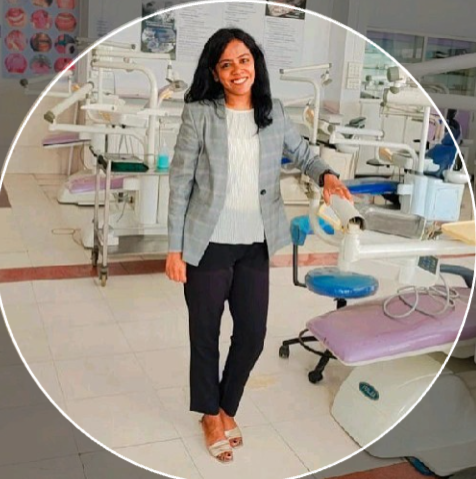
Dr. Shraddha Supnekar interprets CBCT, OPG, and cross-sectional maxillofacial imaging — seven years of high-volume clinical reporting and academic practice, paired with applied volumetric and AI-assisted analysis.
DegreeMDS, Oral Medicine & Radiology — University Topper, Bronze Medallist, MUHS
LicenseDHA Specialist Eligibility, Dental Radiology (Dubai Health Authority)
Experience7+ years post-MDS clinical & academic practice — Assistant to Associate Professor, M.A. Rangoonwala College of Dental Sciences & Research Centre, Pune (2018–2025)
ReportingHigh-volume CBCT & OPG reporting across NewTom/NNT Viewer, Kodak Carestream, and Vatech EzDent/EzImplant platforms
ResearchCNN/ResNet-50 model for dentigerous cyst detection on CBCT; ITK-SNAP–based volumetric segmentation of maxillofacial sinus anatomy
§ 02 · Cases
07 CASE STUDIES
Click a scan to inspect it.
Click :: inspect Click outside :: close
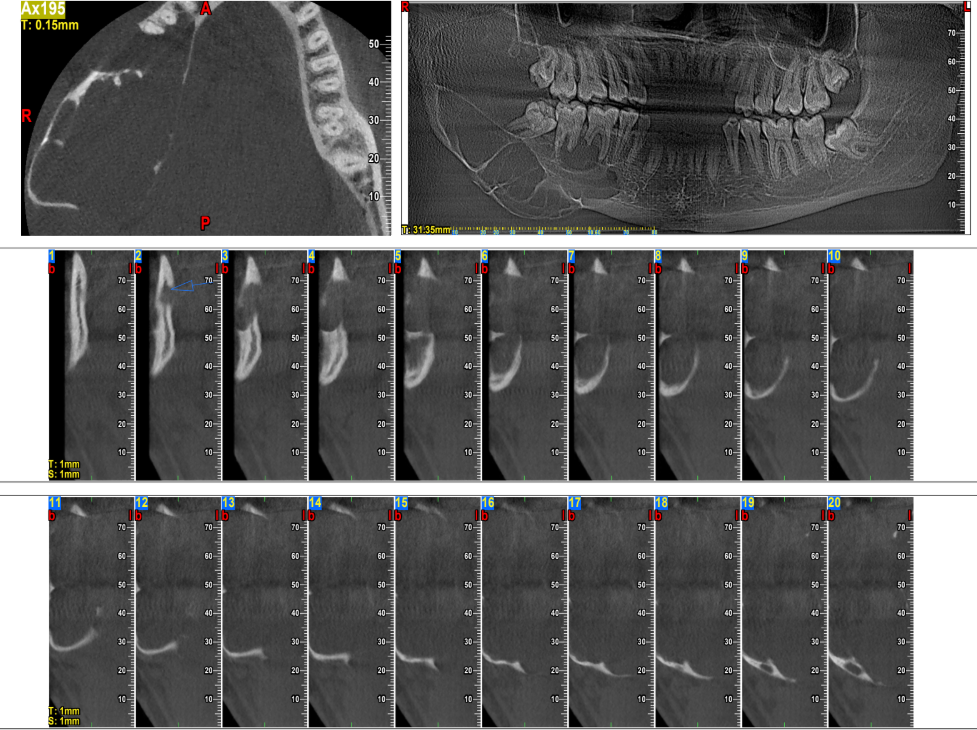
01 · Dentigerous Cyst — Case 1CBCT
SLICE 124/312
ORIENTATION AX
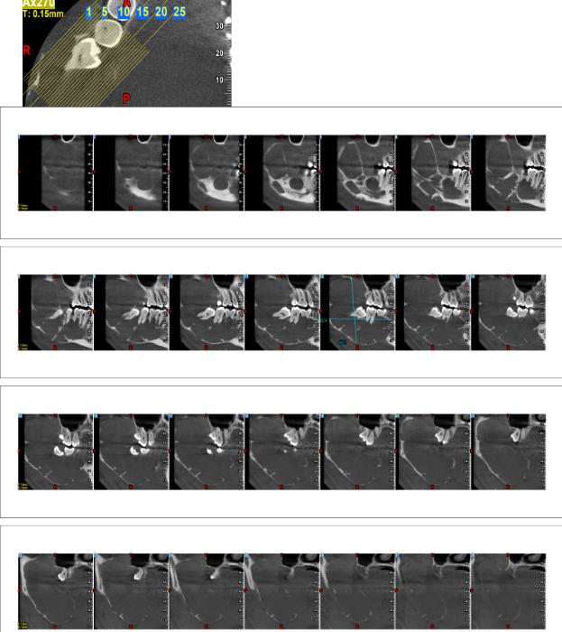
Dentigerous Cyst
Well-defined unilocular radiolucency associated with an impacted tooth.
Modality CBCTFinding Unilocular
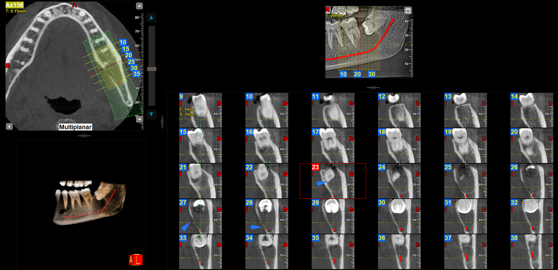
02 · Dentigerous Cyst — Case 2CBCT · 3D
SLICE 156/312
ORIENTATION SAG
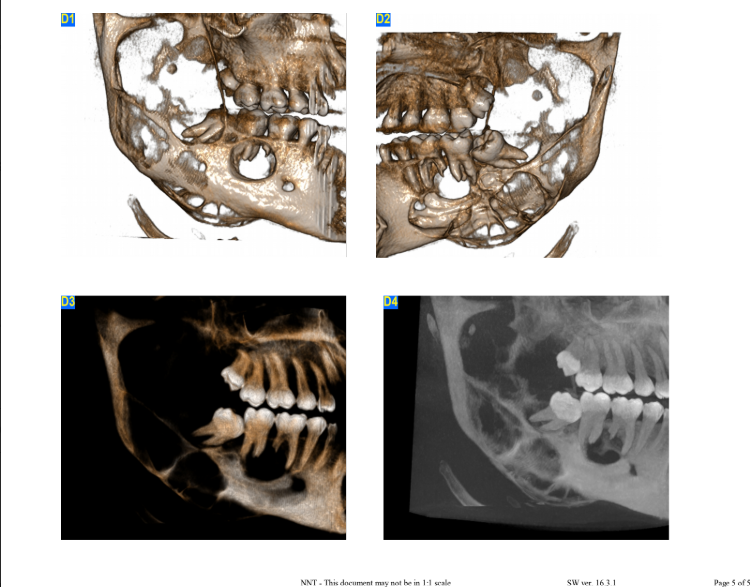
Dentigerous Cyst — Multiplanar
Impacted mandibular tooth with pericoronal radiolucency and cortical expansion, in close proximity to the inferior alveolar canal.
Modality CBCT · MP + 3DFinding Canal proximity
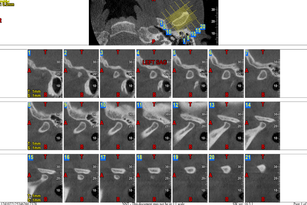
03 · Left TMJ — DegenerativeCBCT · SAGITTAL
SLICE 089/240
ORIENTATION SAG
Left TMJ — Degenerative Changes
Sagittal series demonstrating condylar flattening, consistent with early degenerative joint changes.
Modality CBCT · SAGRegion Left TMJ
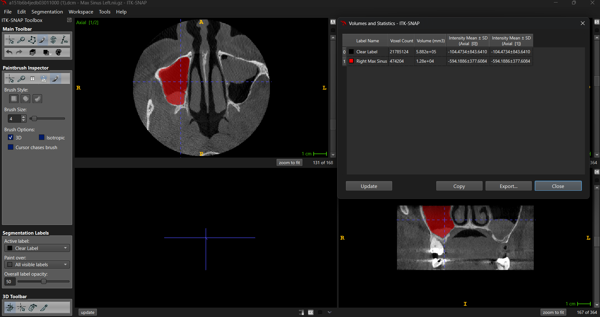
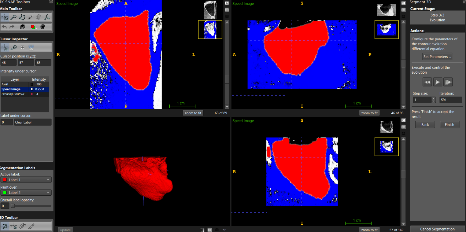
04 · Rt. Maxillary Sinus — Volumetric◉ RESEARCH
METHOD ITK-SNAP
VOXEL 0.15mm³
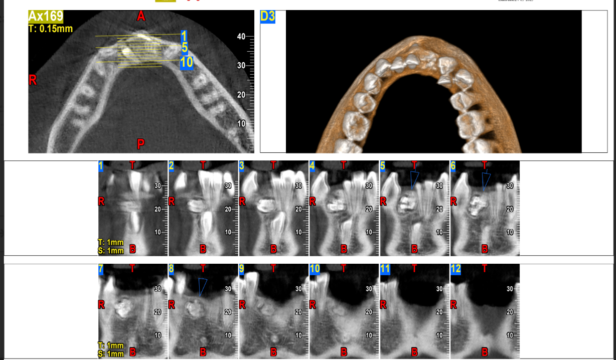
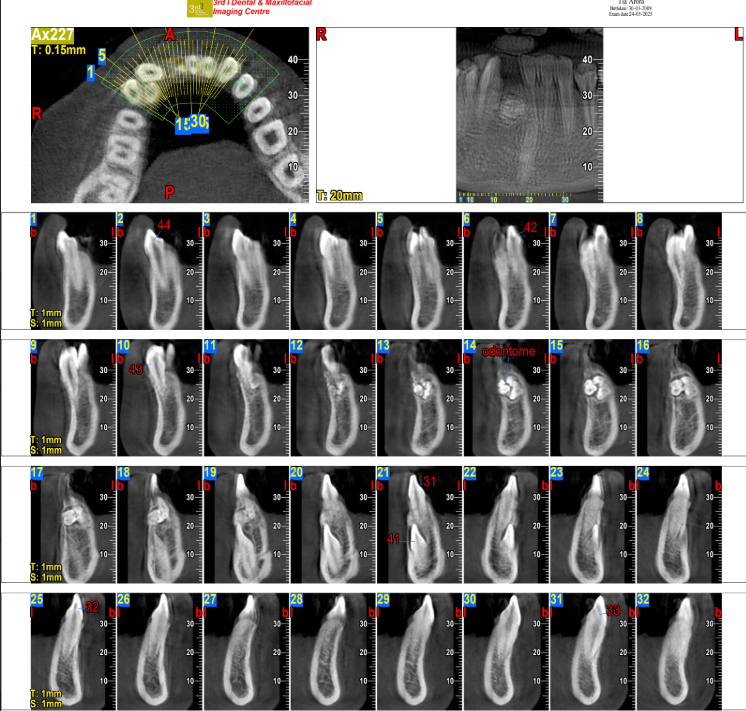
Right Maxillary Sinus — 3D Volumetric Segmentation
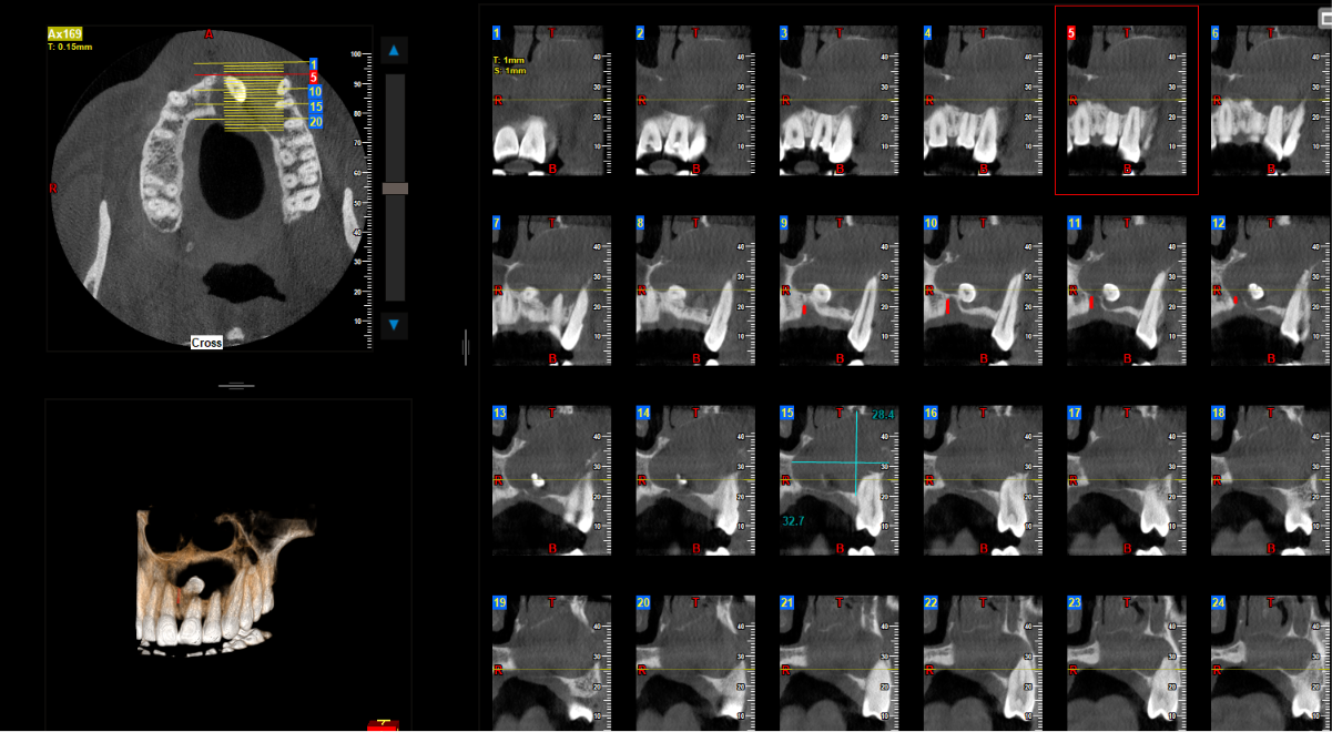
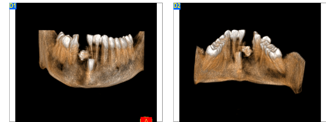
Well-defined mass with tooth-like radiopacities disrupting normal arch continuity — consistent with a compound/complex odontoma.
Modality CBCT · 3D + MPFinding Compound
06 · AmeloblastomaCBCT · 3D + CROSS
SLICE 175/312
MULTILOCULAR ◉
Ameloblastoma
Multilocular, expansile radiolucent lesion of the posterior mandible with cortical thinning — findings consistent with ameloblastoma.
Modality CBCT · 3D + CSFinding Multilocular
07 · Fibro-Osseous LesionCBCT · CORONAL + 3D · BILATERAL
SLICE 145/312
ORIENTATION COR
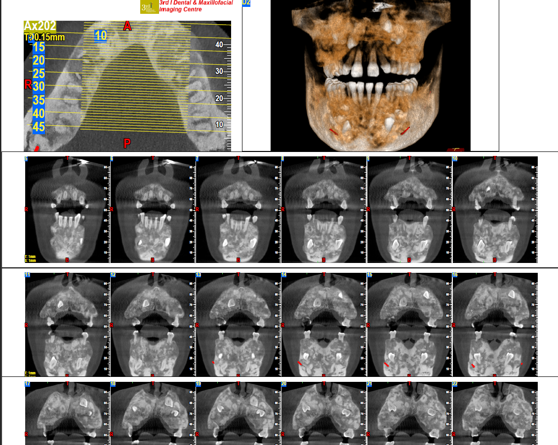
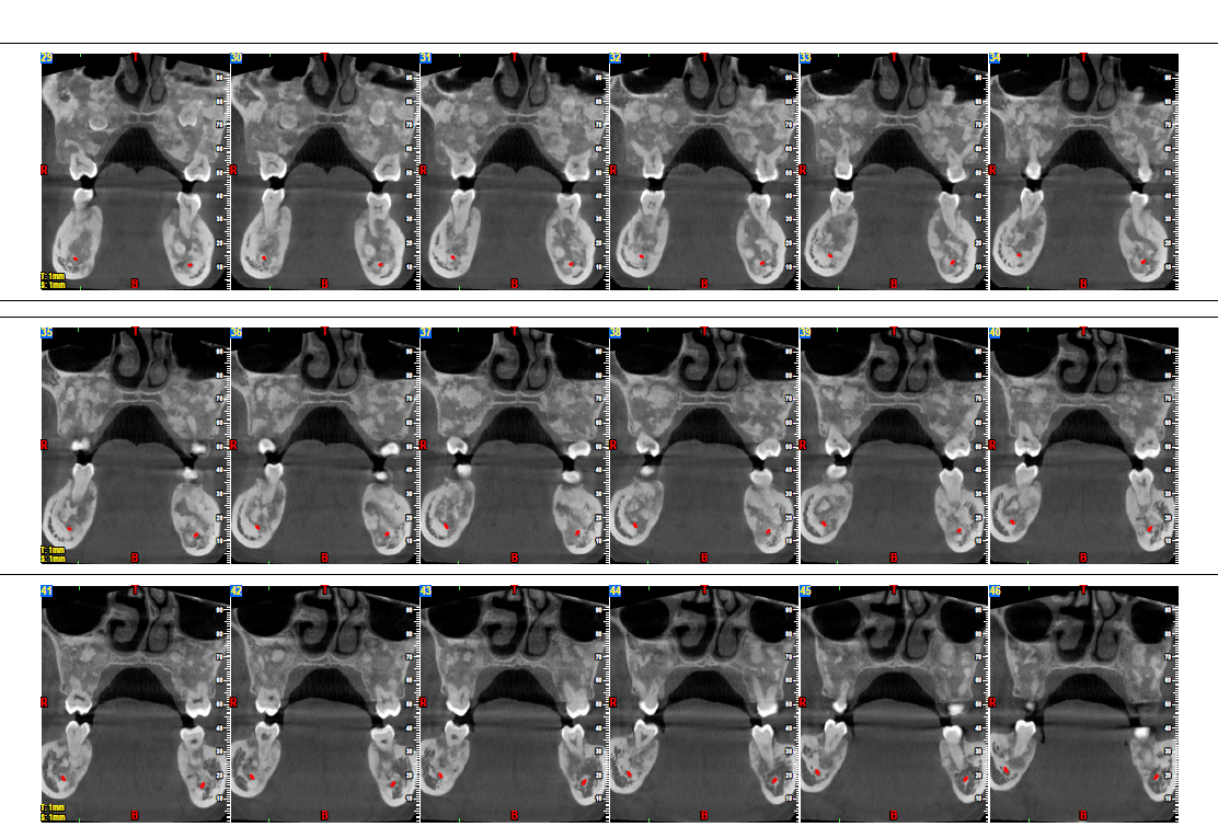
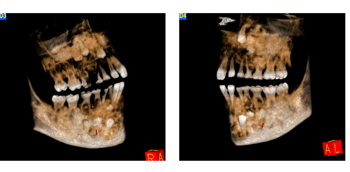
Fibro-Osseous Lesion
Coronal and 3D reconstructions showing bilateral mixed radiopaque–radiolucent lesions with a ground-glass internal pattern — consistent with a fibro-osseous process such as fibrous dysplasia.
Modality CBCT · CORONAL + 3D
Pattern Bilateral ground-glass
Impression Fibrous dysplasia
R.
§ 03 · Research
Applied AI + volumetric analysis
Beyond routine reporting: applied AI and volumetric methods.
01
Deep Learning · In Pipeline
CNN / ResNet-50 detection model.
A deep learning model in development to detect dentigerous cysts on CBCT imaging — built on DICOM-to-PNG extraction with bone-window radiological processing and patient-level train/validation/test splitting.
◉ PIPELINE · TRAIN/VAL/TEST
Train 70%
Val 20%
Test 10%
▸ DICOM → PNG extraction
▸ bone-window preprocessing
▸ patient-level splits
▸ ResNet-50 backbone
02
Volumetric Segmentation
ITK-SNAP segmentation pipeline.
Semi-automated segmentation of maxillofacial sinus anatomy — producing quantitative volume and voxel-level statistics rather than qualitative description alone.
◉ VOLUME · QUANTITATIVE
1.28×10⁴
mm³ segmented volume
Voxel
0.15mm³
Method
Semi-auto
Region
Rt. Maxillary
03
Publications
Thirteen peer-reviewed papers.
Spanning oral medicine, radiology, and the application of AI and deep learning to maxillofacial imaging.
◉ INDEXED · 2020 → 2022
10 OF 13 SOURCED
01
Localized ridge augmentation with mandibular block autograft and guided bone regeneration: a case report
J. Surgical Case Reports & Images · 2022 · Crossref, Publons
02
CBCT report of 3 cases of cysts and its radiographic presentations
Intl. J. Scientific Development & Research · 2022 · Index Copernicus, Google Scholar
03
Oral lichen planus and its variants — case series
Intl. J. Scientific Development & Research · 2022 · Index Copernicus, Google Scholar
04
A rare occurrence of dens invaginatus with mandibular third molar and its CBCT evaluation
European J. Pharmaceutical & Medical Research · 2022 · Google Scholar, Index Copernicus
05
Recurrent aphthous stomatitis — a review
Intl. J. Scientific Development & Research · 2022 · Index Copernicus, Google Scholar
06
Audiometry analysis for the assessment of hearing deficit in patients with oral submucous fibrosis
J. Clinical & Diagnostic Research · 2021 · Index Copernicus, Crossref, Citefactor
07
Position of posterior superior alveolar artery in relation to the maxillary sinus using CBCT in an Indian sub-population
J. Oral Medicine & Oral Surgery · 2021 · Scopus
08
Attitude of IV BDS and intern students towards pursuing a PG course in oral medicine & radiology
Intl. J. Innovative Science & Research Tech. · 2020 · Publons
09
Exploring Nikolsky's sign
IOSR J. Dental & Medical Sciences · 2020 · PubMed
10
Synovial chondromatosis of left TMJ — a rare case with typical imaging feature
European J. Pharmaceutical & Medical Research · 2020 · Index Copernicus
11
Additional publication — details pending
12
Additional publication — details pending
13
Additional publication — details pending
§ 04 · Availability
Current lanes · Open to
Open across three lanes.
OpenL-01
Clinical & Teleradiology
Specialist CBCT/OPG reporting roles, subject to facility DHA sponsorship activating full license status.
OpenL-02
Academic Faculty
Faculty positions in oral & maxillofacial radiology, radiographic interpretation, and imaging sciences.
OpenL-03
Research & AI
Applied deep learning and volumetric imaging projects in maxillofacial diagnostics — collaborative or contract-based.
§ 05 · Portal
Response within 48h
Send a case. Drop the volume.
DICOM Case
PDF Second Opinion
General Inquiry
◉ Response window
< 48 hrs
Every case gets a personal reply — not an autoresponder.
◉ What I need from you
▸Patient age & relevant clinical history
▸Specific focus area or question
▸Prior reports, if any
▸I'll reply with a secure link for the full DICOM stack
◉ Confidentiality
All uploads are treated as protected clinical material. De-identify where practical; identifiers are never retained beyond the reporting window.